
News
- Home
- Love to Know What is DICOM GSDF in Diagnostic Accuracy

Love to Know What is DICOM GSDF in Diagnostic Accuracy
- October 16, 2025
- Sam Lee
The Unseen Hero of Medical Imaging | Unraveling DICOM GSDF
In the intricate world of medical diagnostics, where every pixel can hold the key to a correct diagnosis, the clarity and consistency of images are paramount. Behind the scenes, ensuring this critical visual fidelity is an often-overlooked hero: the DICOM Grayscale Standard Display Function (GSDF). If you’ve ever wondered what is DICOM GSDF and why it’s so vital, or sought a deep dive into DICOM Part 14 explained, you’ve come to the right place. This comprehensive guide will illuminate the science and necessity of the DICOM grayscale standard and introduce you to solutions like PerfectLum that bring its benefits to life.
The Genesis of a Standard: Why We Needed DICOM GSDF
Before the advent of the DICOM GSDF, diagnostic imaging was a wild west of visual inconsistency. Different monitors, graphics cards, and display settings meant that the exact same medical image could appear vastly different from one workstation to another, or even on the same monitor at different times. Imagine a radiologist reviewing an X-ray on a bright, high-contrast screen, only for their colleague to see it later on a dimmer, less calibrated display, potentially missing a subtle anomaly. The consequences of such discrepancies in medical diagnoses are, quite frankly, terrifying.
Recognizing this critical flaw, the medical imaging community, under the umbrella of the National Electrical Manufacturers Association (NEMA) and the American College of Radiology (ACR), developed the Digital Imaging and Communications in Medicine (DICOM) standard. DICOM GSDF is a comprehensive set of rules that defines how medical images and related information are handled, stored, printed, and transmitted. DICOM Part 14, specifically, addresses the Grayscale Standard Display Function (GSDF), providing a standardized way to ensure that grayscale images are displayed consistently across various devices.
What is DICOM GSDF? The Science of Consistent Vision
At its core, the DICOM GSDF is a lookup table (LUT) or a mathematical function that maps Digital Driving Levels (DDLs) – the numerical values representing grayscale pixel intensities – to a specific luminance output on a display device. But it’s not just any mapping; it’s a perceptually linearized mapping.
To understand this, consider how the human visual system perceives changes in brightness. We are not linearly sensitive to light. We perceive differences more acutely in darker tones than in brighter ones. A small change in luminance in a dark area of an image is far more noticeable than the same absolute change in a bright area. Without a standardized function, a display might distribute its brightness levels uniformly, leading to a loss of discernible detail in critical dark regions of medical images.
The DICOM GSDF takes this physiological fact into account. It’s based on extensive psychophysical research into human contrast perception. The function ensures that, for every step in the DDL, the perceived change in brightness is roughly equal. This “perceptual linearization” is crucial because it means:
- Consistent Contrast Perception: Regardless of the display device, a given DDL will always translate to the same perceived brightness difference. This allows radiologists and clinicians to make consistent judgments about image features.
- Optimal Detail Visibility: The DICOM GSDF optimizes the display of detail across the entire grayscale range, particularly enhancing the visibility of subtle nuances in both dark and bright areas of an image, which is vital for detecting pathologies.
- Device Independence: The ultimate goal is that a diagnostically relevant feature, such as a subtle lesion, will appear with the same contrast and clarity whether viewed on a high-end diagnostic monitor in one hospital or a different calibrated monitor in another.
DICOM Part 14 Explained: The Technical Deep Dive
DICOM Part 14 explained specifies the actual mathematical function and the conditions under which it should be applied. It defines the relationship between the input DDL (representing the original image data) and the output luminance (the light emitted by the display). The standard mandates that medical displays used for diagnostic purposes must be calibrated to conform to this GSDF.
Here’s a simplified breakdown of the technical aspects:
- Luminance Range: The GSDF is defined over a specific range of luminances, typically from 0.05 cd/m² (candelas per square meter) to 4000 cd/m². This range encompasses the typical operating brightness of diagnostic displays.
- Input Values (P-Values): The standard defines a range of P-Values (Presentation Values) from 0 to 4095. These are essentially normalized DDLs.
- The Function: The core of Part 14 is a complex logarithmic function that maps these P-Values to a specific output luminance (L). The function is designed to produce a luminance response that closely matches the human visual system’s perception of contrast, ensuring that equally spaced P-Values result in equally perceived brightness steps.
- Calibration: For a display to be DICOM Part 14 compliant, it must be calibrated so that its actual luminance output, for each input DDL, closely matches the luminance prescribed by the GSDF. This is typically done using specialized software and a photometer (a device to measure light intensity).
- Ambient Light: While the GSDF dictates the display’s output, it’s also crucial to consider the ambient lighting conditions in the viewing environment. Excessively bright ambient light can wash out details and negate the benefits of GSDF calibration. DICOM provides recommendations for viewing environments to minimize these effects.
The Critical Role of the DICOM Grayscale Standard in Diagnostic Accuracy
The impact of the DICOM grayscale standard on diagnostic accuracy cannot be overstated. Consider these scenarios:
- Subtle Lesions: Many early-stage cancers or microfractures appear as very subtle changes in grayscale intensity. Without DICOM GSDF, these critical details could be lost in inconsistent display rendering, leading to missed diagnoses.
- Comparative Analysis: Radiologists often compare current images with previous ones to track disease progression or healing. GSDF ensures that these comparisons are valid, as the images are displayed under a consistent perceptual standard.
- Multi-Disciplinary Teams: In a hospital, various specialists – radiologists, oncologists, surgeons – may view the same image on different displays. DICOM GSDF guarantees that they are all seeing the same visual information, facilitating accurate collaboration and treatment planning.
- Legal Implications: In a medical-legal context, demonstrating that diagnostic images were reviewed on DICOM Part 14 compliant displays can be crucial for defending diagnostic decisions.
Ultimately, the DICOM GSDF transforms arbitrary pixel values into a visually consistent and diagnostically reliable representation of the patient’s anatomy, enhancing the confidence and precision of medical professionals.
The Imperative of Calibration and QA: Why PerfectLum is Indispensable
While the DICOM GSDF provides the foundational standard, simply having a “DICOM compliant” monitor isn’t enough. Displays drift over time due to factors like backlight aging and usage. This drift can pull them out of GSDF compliance, reintroducing the very inconsistencies the standard sought to eliminate.
This is where dedicated calibration and quality assurance (QA) software becomes indispensable. This is where solutions like PerfectLum shine.
PerfectLum is not just a calibration tool; it’s a comprehensive QA solution designed specifically for medical displays. Here’s how it ensures that the promise of DICOM Part 14 GSDF is consistently delivered:
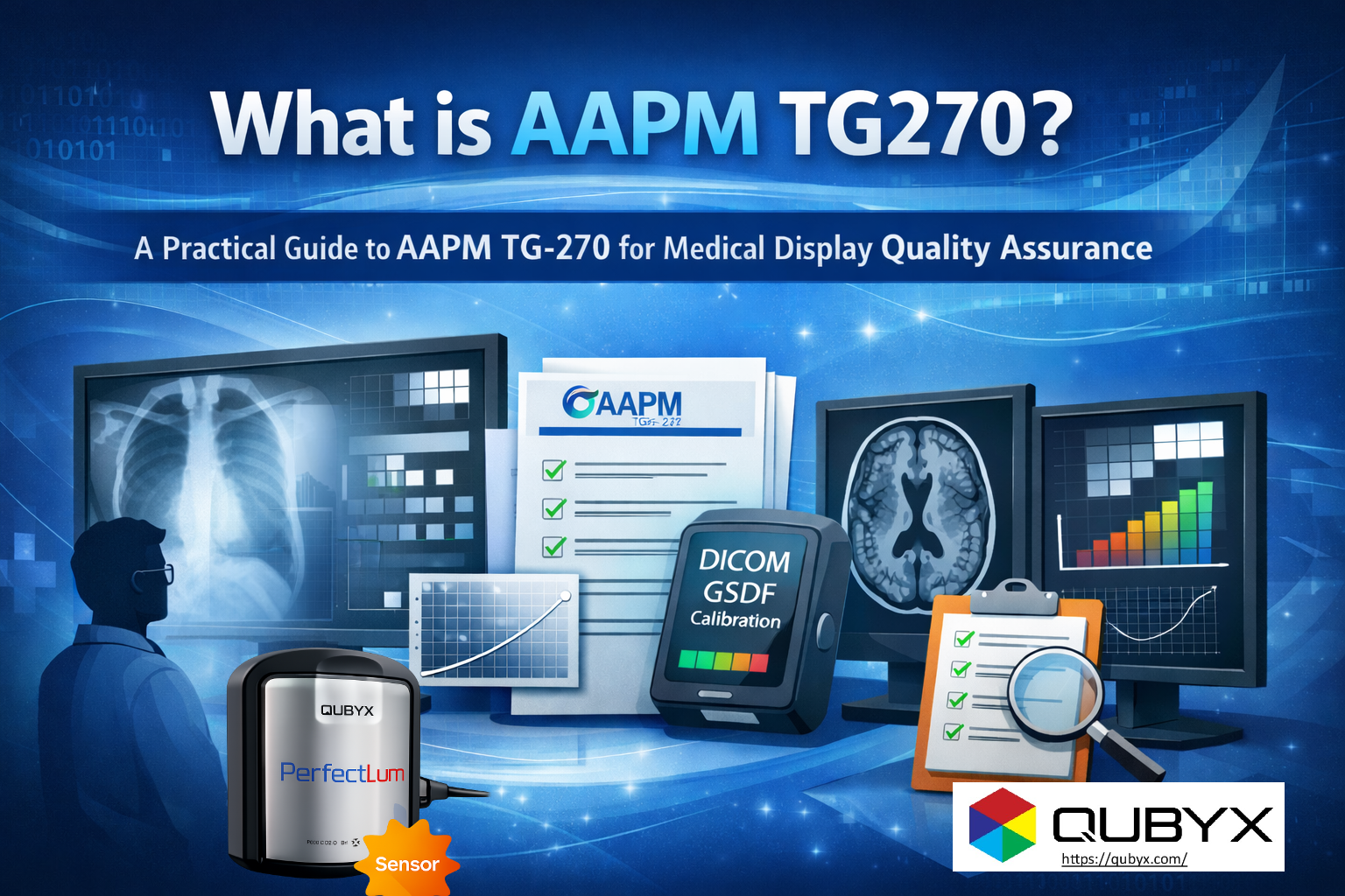
- Automated DICOM Part 14 Calibration: PerfectLum automatically calibrates medical displays to the DICOM GSDF curve. It measures the display’s luminance output at various grayscale levels and adjusts the video card’s LUT to precisely match the GSDF. This ensures perceptual linearity and consistent brightness.
- Ongoing QA and Monitoring: Calibration is not a one-time event. PerfectLum enables regular and automated QA testing, monitoring the display’s performance against critical parameters like luminance, contrast, uniformity, and resolution. It detects drift and prompts re-calibration when necessary.
- Compliance with International Standards: Beyond DICOM Part 14, PerfectLum helps maintain compliance with other critical medical imaging standards, such as those from the American Association of Physicists in Medicine (AAPM) Task Group 18 (TG18) and various national guidelines.
- Comprehensive Reporting: PerfectLum generates detailed reports on calibration and QA tests, providing auditable documentation of display performance. This is crucial for regulatory compliance and accreditation purposes.
- Remote Management: For larger healthcare facilities, PerfectLum offers centralized management capabilities, allowing administrators to monitor and manage multiple displays across different departments or even locations from a single console. This ensures consistent quality across the entire enterprise.
- User-Friendly Interface: Despite its sophisticated underlying technology, PerfectLum is designed to be user-friendly, making it accessible to a wide range of users, from IT professionals to medical physicists and radiologists.
By employing PerfectLum, healthcare providers can move beyond simply understanding what is DICOM GSDF to actively implementing and maintaining its critical benefits. It bridges the gap between the theoretical standard and the practical reality of day-to-day diagnostic imaging, ensuring that every image viewed is of the highest possible quality and consistency.
The Future of Visual Fidelity in Medical Imaging
As medical imaging continues to evolve with higher resolutions, 3D rendering, and advanced visualization techniques, the fundamental principles of the DICOM GSDF remain as relevant as ever. The need for consistent, accurate, and perceptually linearized grayscale displays is not diminishing; it’s intensifying.
The integration of AI and machine learning into diagnostics further underscores this need. AI algorithms trained on images displayed according to the GSDF will perform optimally when presented with similarly calibrated images in clinical practice. Any deviation could introduce errors or reduce the efficacy of these advanced tools.
Investing in robust display QA solutions like PerfectLum is not merely a compliance exercise; it’s an investment in patient safety, diagnostic confidence, and the efficiency of healthcare delivery. It ensures that the “unseen hero” of the DICOM Grayscale Standard Display Function continues to perform its vital role, empowering clinicians to make the best possible decisions based on the most accurate visual information.
Conclusion
The DICOM Grayscale Standard Display Function, detailed in DICOM Part 14 explained, is a cornerstone of modern medical imaging. It’s the invisible force that standardizes how we perceive critical diagnostic information, moving medical imaging from a subjective art to a precise science. Understanding what is DICOM GSDF reveals its profound impact on patient care. And in the ongoing quest for absolute visual fidelity, solutions like PerfectLum are essential tools, transforming the abstract concept of a standard into a tangible, consistently high-quality reality on every diagnostic screen. For healthcare institutions committed to excellence, integrating DICOM Part 14 compliance with continuous QA is not an option, but a necessity.
Tags:
what is DICOM GSDF, DICOM Part 14 explained, DICOM grayscale standard, PerfectLum, medical display calibration, GSDF compliance, diagnostic accuracy, DICOM standard, medical imaging QA, grayscale standard display function, QYBYX,
Related Posts

- February 24, 2026
- News
What is AAPM TG270? A Practical Guide to AAPM TG-270

- February 24, 2026
- News
How to Calibrate a Display to DICOM GSDF with PerfectLum

- February 23, 2026
- News
Why Calibrate a Display to DICOM? And the Role