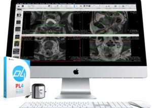
In the healthcare niche, the quality of the diagnostic monitor holds a great significance to produce accurate biomedical images, image-related information, and further diagnosis based on model output. Where the traditional computer monitor fails to capture the core details, DICOM calibration enables medical equipment to produce images more clearly and precisely.

DICOM stands for Digital Imaging and Communications. DICOM ensures the standardized regulation for medical imaging color prospects for GSDF (Grayscale Standard Display Function) as per the (NEMA) recent guidelines to achieve better and correct diagnosis results. National Electrical Manufacturers Association (NEMA) and the American College of Radiology (ACR) in joint collaboration have set out guidelines for the global standard of medical imaging practices for the calibration of grayscale. DICOM aims to ensure compliance with the DICOM GSDF (Grayscale Standard Display Function) standard. In easy words, it defines the process of how grayscale images must be produced and showcased on medical displays.
DICOM shares the guidelines for handling, storing, sharing, and printing of medical imaging. DICOM calibration software holds the capability to read digital images that come from a wide range of modalities, including ultrasound, CTs, MRIs, x-rays, and mammographies, among many other similar complex images formats.
Understanding DICOM
In order to understand the significance of DICOM, we need to go back to the earlier traditional grayscale imagining screens and its associated drawbacks. So the basic distinctions are quality, usage, and production capability. Where the computer monitor was restricted in its use and compatibility, the technology-driven DICOM software supports digital medical images from a variety of capture modalities with ease. What’s more, it offers global access reach with multi workstation capability.
Area of application
With the proven effectiveness of the DICOM Standard, The DICOM calibration has come out as the world leader to define the standard for image handling in the medical niche. DICOM shares a wide area of usages in different processes in hospitals, clinical departments, and medical institutions.
DICOM Service usually shares the five general application areas.
- Network image management
- Network image interpretation management
- Network print management
- Imaging procedure management
- Off-line storage media management
If we speak more specifically to the medical process then it would include the departments:
- Surgery
- Radiology
- Digital Imaging and Communications in Medicine
- Cardiology
- Radiotherapy
- Oncology
- Pathology
- Dentistry
- Ophthalmology
- Veterinary
- Neurology
- Pneumology
- Oncology
Features
- EASY to use & install
- Validated and certified
- Complete REPORTING and history database
- Built-in Quality Management system for NYPDM and NYCPDM
- Remote QA SERVER
- FAST calibration
Benefits
DICOM calibration helps medical practitioners in multiple ways to boost their research and medical practices. Here is how it helps in deriving better and improved results:
Increasing Luminance: The qualities of DICOM software can be defined in one simple sentence that it helps produce images that can be visible to naked eyes. DICOM software fortifies standardized luminance all over the image to provide detailed information of images.
Consistency and Accuracy: Grayscale is limited in quality to catch every small element in an image. Without the calibration, images like X-rays are fully unrecognizable to the human eye to detect defects. DICOM removes all such areas!
Likewise, it supports you to produce images recurrently without investing more time and effort.
Image Sharing: This is the most impressive part of DICOM calibration. You can share, and view the image in multiple screens without any location restriction to get the same results without losing a single bit of image detailing.


